Ultramarine Blue: Its Color Comes Mostly From Caged Sulfur
Tricks of Ear and Eye Published
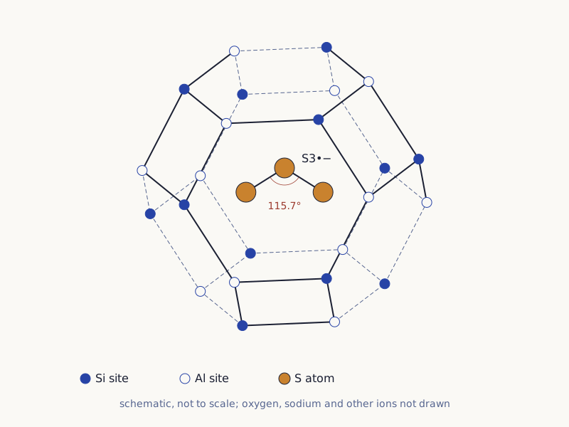
The bent chain of three sulfur atoms in the cage in Figure 1 below is mostly what makes ultramarine blue. The cage is part of lazurite, the blue mineral in lapis lazuli. The trapped sulfur group, called the trisulfur radical, is thought to be mainly responsible for the blue [1]. Chemists made a factory version in the 1820s: Hamerton and colleagues (2013) recount a French prize offered in 1824 and given to Guimet in 1828 [2].
Wide drawing on a small screen: swipe sideways, or open it full size.
Figure 1. One sodalite-type cage with the trisulfur radical inside, drawn as a schematic; oxygen and the other ions are left out.
Diagram: Tricks of Ear and Eye, CC BY 4.0
Full credit line to copy
"Sodalite-type cage with a trisulfur radical, schematic" by Tricks of Ear and Eye, https://tricksofearandeye.com/color/ultramarine/, licensed under CC BY 4.0, https://creativecommons.org/licenses/by/4.0/
Data: Cage description from Chukanov, N. V., & Aksenov, S. M. (2024), Int. J. Mol. Sci. 25: 10218, CC BY 4.0; S3 angle from Chivers, T., & Oakley, R. T. (2023), Molecules 28: 5654, CC BY 4.0
What makes ultramarine blue?
Count the corners in Figure 1. There are 24, and filled and open circles take turns all the way round. They stand for silicon and aluminum sites, which alternate in this kind of mineral frame [3]. Chukanov and Aksenov (2024) give the cage the symbol [4668]: six rings of four sites and eight rings of six [3].
The drawing check (CSV) (where the data come from and its license, JSON) confirms that Figure 1 has exactly those rings, and that each of its 36 edges joins a silicon site to an aluminum site.
Lazurite has the same frame as the mineral sodalite, and its central holes are called β-cages [1]. Several ions and small sulfur groups have been found trapped in them. One is S3•−: three linked sulfur atoms with an extra negative charge and one unpaired electron, shown by the dot. González-Cabrera and colleagues (2022) write that it is "considered to be predominantly responsible" for the blue [1].
Chukanov and Aksenov go further. They write that spectra led to the conclusion that S3•− causes the deep blue of lazurite and ultramarine, and that "numerous subsequent studies confirmed this conclusion" [3]. Very little of it is needed. They report that minerals of this group with about 0.01 S3•− per formula unit already have an intense blue color [3].
The cage matters too. In a liquid, these sulfur groups react readily with oxygen from the air, but the small ones can last when trapped in the mineral frame (Chivers & Oakley, 2023) [4]. The same authors list S2•− as the color source in yellow ultramarine, S3•− in blue, and both together in green [4].
Figure 1 bends the chain at 115.7 degrees. That is the angle Chivers and Oakley calculated for the ion on its own, not one measured inside a cage [4].
Which light does the radical absorb?
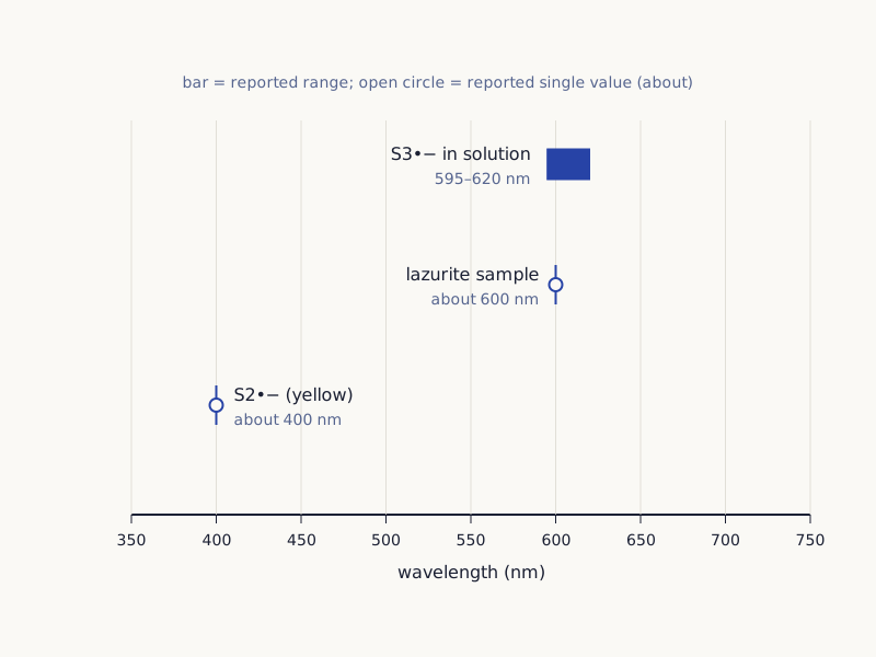
Look at the bar at the top of Figure 2. S3•− absorbs most strongly between 595 and 620 nanometers, and the exact spot depends on the liquid around it [3]. In a dark blue lazurite sample, the broad absorption peak sits at about 600 nanometers [3]. The yellow S2•− absorbs most near 400 nanometers instead [3].
Wide drawing on a small screen: swipe sideways, or open it full size.
Figure 2. Where the sulfur color centers absorb most strongly; positions only, with no curve shapes or colors drawn.
Chart: Tricks of Ear and Eye, CC BY 4.0
Full credit line to copy
"Absorption positions of the sulfur color centers in ultramarine" by Tricks of Ear and Eye, https://tricksofearandeye.com/color/ultramarine/, licensed under CC BY 4.0, https://creativecommons.org/licenses/by/4.0/
Data: Absorption positions from Chukanov, N. V., & Aksenov, S. M. (2024), Int. J. Mol. Sci. 25: 10218, CC BY 4.0
The color-center table (CSV) (where the data come from and its license, JSON) Data: Values from Chukanov & Aksenov 2024 (CC BY 4.0) and Chivers & Oakley 2023 (CC BY 4.0), read in full, and from the abstract of Eaton, Mitchell & Eaton 2025; one ratio computed by Tricks of Ear and Eye lists these values with their sources and the calculated shape of the chain. The papers read for this page link the blue to this absorption. They do not model how the eye responds to it.
Is ultramarine the same as lapis lazuli?
No. Lapis lazuli is a rock, and lazurite is the blue mineral in it [1]. The file page for the stone in Figure 3 describes it as lapis lazuli with pyrite, from Afghanistan. Pyrite, an iron sulfide, is one of the impurities González-Cabrera and colleagues found in lower-quality natural pigment [1].
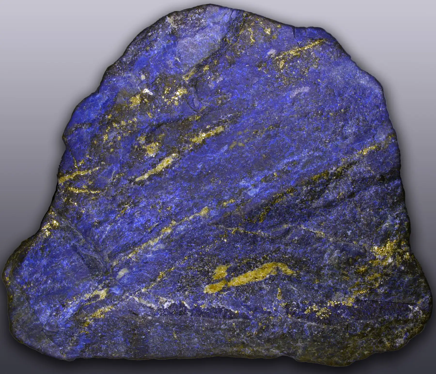
Figure 3. Lapis lazuli with pyrite, from Afghanistan, photographed in 2008.
"Lapis lazuli with pyrite, Afghanistan" by Hannes Grobe — https://commons.wikimedia.org/wiki/File:Lapis-lazuli_hg.jpg — CC BY-SA 2.5 (https://creativecommons.org/licenses/by-sa/2.5/)
Natural ultramarine is the blue part taken out of that rock. The same team mapped where the lazurite signal showed up in each sample [1]. It covered 85% of the mapped pixels in a synthetic pigment but only 7% in "ultramarine ash," the by-product left once the blue is removed [1]. One of their natural samples had been purified by the traditional method described by Cennino Cennini; this page gives no steps.
The name comes from the stone's journey. Kraft (2008) notes that lapis lazuli reached Europe from Asia by sea, and that ultramarine means "beyond the sea" [6].
Natural and synthetic
Natural and factory-made ultramarine share the same blue source. According to their abstract, Eaton and colleagues (2025) found that the blue of lazurite is due to the same S3− radical as in synthetic ultramarine blue [7].
The amounts differ. The abstract counts the radicals as spins per gram: 3 × 10²⁰ to 5 × 10²⁰ in three commercial synthetic samples, and 6 × 10¹⁸ to 2.9 × 10¹⁹ in lapis lazuli from four places [7]. That is a factor of about 10 to 83, computed for this page. Whole rock and bought pigment are different kinds of sample, so the factor is a scale aid, not a quality ranking.
Heat leaves its own mark. Gambardella and colleagues (2020) heated lapis lazuli before taking out the pigment [8]. The sulfur signal they link to the trisulfur radical grew, and the blue deepened [8]. They found the same marker in five old paint samples [8].
A 1998 study by Gobeltz and colleagues looks at how the color centers get into the cage during manufacture. It is listed here by title only, because this site could not read it [5].
Hamerton and colleagues (2013) note that in some religious paintings, most of the blue is "a lesser pigment, such as azurite," while the robes of Mary, and sometimes Peter, are ultramarine [2]. This site has not read a pigment study of the painting in Figure 4. So this page makes no claim about which blue it contains.
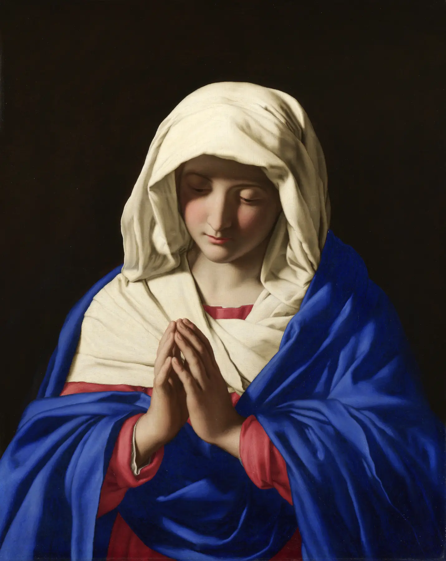
Figure 4. "The Virgin in Prayer" by Sassoferrato, painted between 1640 and 1650, National Gallery, London (NG200); no pigment analysis of it was read for this page.
"The Virgin in Prayer" by Giovanni Battista Salvi da Sassoferrato, public domain
Full credit line to copy
"The Virgin in Prayer" by Giovanni Battista Salvi da Sassoferrato — https://commons.wikimedia.org/wiki/File:Sassoferrato_-_Jungfrun_i_b%C3%B6n.jpg — Public domain (Giovanni Battista Salvi da Sassoferrato died 1685; public domain in the US (published before 1931) and in the EU (author died more than 70 years ago); PD-Art faithful reproduction (photograph from the Web Gallery of Art; National Gallery, London, NG200, as recorded on the Commons file page))
A prize for a recipe
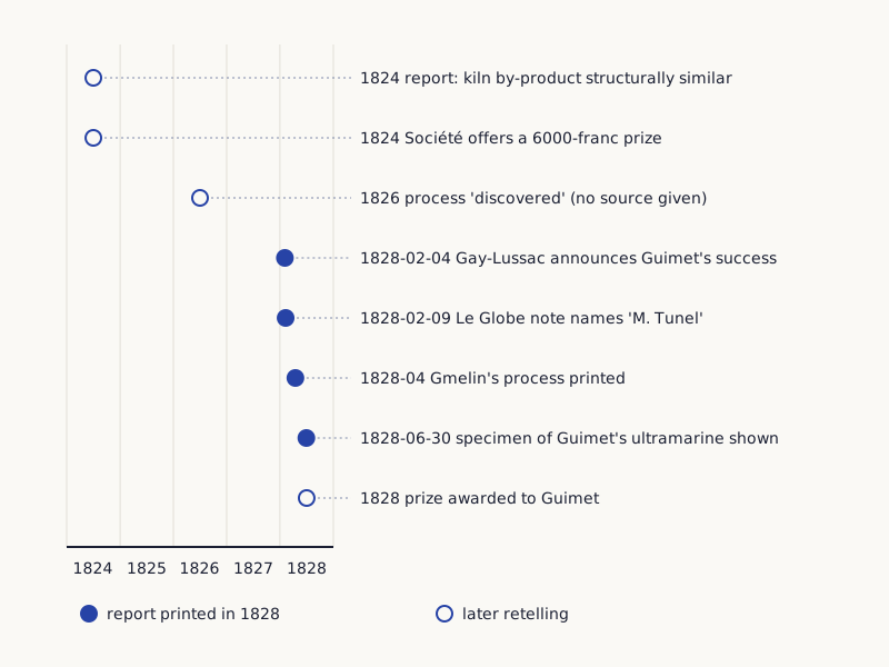
In Figure 5, filled circles are reports printed in 1828. Open circles are later retellings. The account of the prize is one of those retellings. Hamerton and colleagues (2013) take it from a 1993 article in the magazine Chemistry in Britain [2].
By their account, a French chemist they call Vacquelin reported in 1824 that a by-product of glass making in soda kilns was structurally similar to natural ultramarine. The Société d'Encouragement pour l'Industrie Nationale then offered 6000 francs for an economical factory method. The German chemist Gmelin claimed the reward. After that dispute, the prize went to Guimet in 1828 [2].
Wide drawing on a small screen: swipe sideways, or open it full size.
Figure 5. Eight dated records of early factory-made ultramarine, from the timeline table on this page.
Chart: Tricks of Ear and Eye, CC BY 4.0
Full credit line to copy
"Records of early factory-made ultramarine, 1824 to 1828" by Tricks of Ear and Eye, https://tricksofearandeye.com/color/ultramarine/, licensed under CC BY 4.0, https://creativecommons.org/licenses/by/4.0/
Data: Rows of the Tricks of Ear and Eye ultramarine timeline table: Hamerton, Tedaldi & Eastaugh 2013 (CC BY 4.0); The Philosophical Magazine 1828, volumes 3 and 4 (public domain); Kraft 2008
What the 1828 journals printed
The Philosophical Magazine for 1828 lets parts of this story be checked. It reports a sitting of the Paris Academy of Sciences on 4 February 1828, where Gay-Lussac announced that Guimet, of the gunpowder and saltpetre works, had succeeded in making ultramarine [9].
A note from the newspaper Le Globe of 9 February tells it differently: it names the maker as "M. Tunel, inspector of gunpowder and saltpetre," and says the method was being kept secret [10]. A later note adds that Gmelin's own method had appeared in the April Annales de Chimie [11]. On 30 June, Gay-Lussac showed the Academy a sample of Guimet's ultramarine [9].
This site found no readable copy of the Société's own records. So the prize and its terms rest on Hamerton's retelling. Kraft (2008) gives yet another date: a note says a method was found in 1826, with no source given [6]. A 1966 paper by Plesters, "Ultramarine blue, natural and artificial," could not be read for this page [12].
| Date | Record | Kind of evidence |
|---|---|---|
| 1824 | Report that a by-product of glass making in soda kilns was structurally similar to natural ultramarine | Later retelling (Hamerton et al., 2013) |
| 1824 | 6000-franc prize offered for an economical factory method | Later retelling (Hamerton et al., 2013) |
| 1826 | A method said to have been found | Later retelling, no source given (Kraft, 2008) |
| 4 February 1828 | Gay-Lussac announces Guimet's success at the Academy of Sciences | Report printed in 1828 |
| 9 February 1828 | Le Globe note names "M. Tunel" and a secret method | Report printed in 1828 |
| April 1828 | Gmelin's method printed in the Annales de Chimie | Report printed in 1828 |
| 30 June 1828 | Sample of Guimet's ultramarine shown to the Academy | Report printed in 1828 (year from the December 1828 issue) |
| 1828 | Prize given to Guimet after Gmelin's claim | Later retelling (Hamerton et al., 2013) |
The timeline table (CSV) (where the data come from and its license, JSON) Data: Hamerton, Tedaldi & Eastaugh 2013, PLoS ONE (CC BY 4.0), retelling Butler & Gash 1993; The Philosophical Magazine and Annals of Philosophy 1828 (public domain); Kraft 2008, Bulletin for the History of Chemistry (one date) has the same rows with full source details.
What the records leave out
Old recipes rarely give the key details. Hamerton's team followed old texts and got cream, grey and pink products before they got a usable blue [2]. They note that the recipes tend to leave out temperatures and times [2]. The old method burns sulfur in a furnace, and sulfur dioxide forms as it does [2]; this page explains the chemistry only and gives no recipe.
Prices are left out on purpose. The franc prices in Hamerton's paper sit in its retelling of the 1993 magazine article [2]. The 1828 Le Globe note quotes a maker's own selling price [10]. Neither is a record of what buyers paid, so this page compares no prices.
Two other blues help place these dates. Prussian blue was first mentioned in the scientific literature in 1710, in a Berlin publication of the Royal Prussian Society of Sciences [6]. Hamerton and colleagues open their paper with Egyptian blue, a pigment made by the ancient Egyptians [2].
The swatch problem
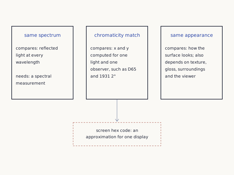
Figure 6 splits "the same color" into three separate claims. That is why this page shows no hex swatch for ultramarine.
Wide drawing on a small screen: swipe sideways, or open it full size.
Figure 6. Three different claims inside "the same color," an editorial distinction drawn for this page rather than a measurement.
Diagram: Tricks of Ear and Eye, CC BY 4.0
Full credit line to copy
"Three meanings of the same color" by Tricks of Ear and Eye, https://tricksofearandeye.com/color/ultramarine/, licensed under CC BY 4.0, https://creativecommons.org/licenses/by/4.0/
| Claim | What is compared |
|---|---|
| Same spectrum | Reflected light at every wavelength |
| Chromaticity match | x and y computed for one light and one observer, such as D65 and the 1931 2° observer |
| Same appearance | How the surface looks, which also depends on texture, gloss, surroundings and the viewer |
| Screen hex code | An approximation for one display |
Even careful color numbers depend on choices. Hamerton and colleagues worked out theirs from measured spectra "under D65 illumination, and the 1931 2° observer" [2]. Those are a standard light and a standard viewer. For the products of their best method, they report chromaticity values, two numbers that place a color on a standard map, of x 0.2945 to 0.3125 and y 0.2219 to 0.2617 [2].
They also warn that the colors in their tables are "merely indicative of hue and not exact representations of the colours achieved" [2]. This site takes the same line. A hex code is a screen approximation. A matching spectrum, a matching chromaticity and a matching look are three different claims.
The checker-shadow entry comes at this gap from the other side. It uses two patches with one gray value in different surroundings. This site's own blue is named after ultramarine, but it is a design choice, not a measurement of the pigment.
Sources
- González-Cabrera, M., Wieland, K., Eitenberger, E., Bleier, A., Brunnbauer, L., Limbeck, A., Hutter, H., Haisch, C., Lendl, B., Domínguez-Vidal, A., & Ayora-Cañada, M. J. (2022). Multisensor hyperspectral imaging approach for the microchemical analysis of ultramarine blue pigments. Scientific Reports 12: 707. Open the sourceRead: full textText license: CC BY 4.0
- Hamerton, I., Tedaldi, L., & Eastaugh, N. (2013). A systematic examination of colour development in synthetic ultramarine according to historical methods. PLoS ONE 8(2): e50364. Open the sourceRead: full text; its history of the 1824 prize is retold from a 1993 magazine articleText license: CC BY 4.0
- Chukanov, N. V., & Aksenov, S. M. (2024). Structural features, chemical diversity, and physical properties of microporous sodalite-type materials: A review. International Journal of Molecular Sciences 25(18): 10218. Open the sourceRead: full textText license: CC BY 4.0
- Chivers, T., & Oakley, R. T. (2023). Structures and spectroscopic properties of polysulfide radical anions: A theoretical perspective. Molecules 28(15): 5654. Open the sourceRead: full textText license: CC BY 4.0
- Gobeltz, N., Demortier, A., Lelieur, J. P., & Duhayon, C. (1998). Encapsulation of the chromophores into the sodalite structure during the synthesis of the blue ultramarine pigment. Journal of the Chemical Society, Faraday Transactions 94: 2257–2260. Open the sourceRead: bibliographic record only; not read
- Kraft, A. (2008). On the discovery and history of Prussian blue. Bulletin for the History of Chemistry 33(2): 61–67. Open the sourceRead: full text; paraphrased
- Eaton, S. S., Mitchell, D. G., & Eaton, G. R. (2025). Electron spin relaxation of S3− in ultramarine blue and lapis lazuli. Applied Magnetic Resonance 56(1–2): 73–90. Open the sourceRead: abstract only
- Gambardella, A. A., Cotte, M., de Nolf, W., et al. (2020). Sulfur K-edge micro- and full-field XANES identify marker for preparation method of ultramarine pigment from lapis lazuli in historical paints. Science Advances 6(18): eaay8782. Open the sourceRead: full text; paraphrased
- Royal Academy of Sciences of Paris: reports of the sittings of 4 February and 30 June 1828. The Philosophical Magazine and Annals of Philosophy, new series, 4 (1828): 301–302 and 456. Open the sourceRead: scanned copy, read for verification
- Manufacture of ultramarine [from Le Globe, 9 February 1828]. The Philosophical Magazine and Annals of Philosophy, new series, 3 (1828): 232. Open the sourceRead: scanned copy, read for verification
- Artificial ultramarine. The Philosophical Magazine and Annals of Philosophy, new series, 4 (1828): 228–229. Open the sourceRead: scanned copy, read for verification
- Plesters, J. (1966). Ultramarine blue, natural and artificial. Studies in Conservation 11: 62–75. Open the sourceRead: bibliographic record only; not read
Sense check:
One step, under a minute
Open this page on two screens, such as a phone and a computer, and compare the robe in Figure 4. Note what differs; neither screen shows the pigment itself. Next: Prussian blue · Checker-shadow illusion
Related entries
Prussian Blue: The Accident Story Rests on a 1731 Account
Color & Pigment
Prussian blue was announced in Berlin in 1710 and a recipe printed in 1724. The tale of a red that turned blue comes from Stahl's 1731 book, as Kraft traces it.
Image credit
"Pigment Berliner Blau" by Saalebaer — https://commons.wikimedia.org/wiki/File:Pigment_Berliner_Blau.JPG — CC0 1.0 (https://creativecommons.org/publicdomain/zero/1.0/)
Egyptian Blue: Under Red Light, It Glows in Infrared
Color & Pigment
Egyptian blue, a copper silicate with an earliest known use of 3300–3200 BCE, emits near-infrared light reported near 910 nm, past the CIE curve's 830 nm end.
Image credit
"Shabti of Yuya" by The Metropolitan Museum of Art — https://commons.wikimedia.org/wiki/File:Shabti_of_Yuya_MET_DT541.jpg — CC0 1.0 (https://creativecommons.org/publicdomain/zero/1.0/)
Cite this page
Tricks of Ear and Eye. "Ultramarine Blue: Its Color Comes Mostly From Caged Sulfur." Tricks of Ear and Eye. Published 2026-10-30. https://tricksofearandeye.com/color/ultramarine/. Accessed on the date you read it.