Mantis Shrimp Eyes: A Tested Species Told Hues Apart Poorly
Tricks of Ear and Eye Published
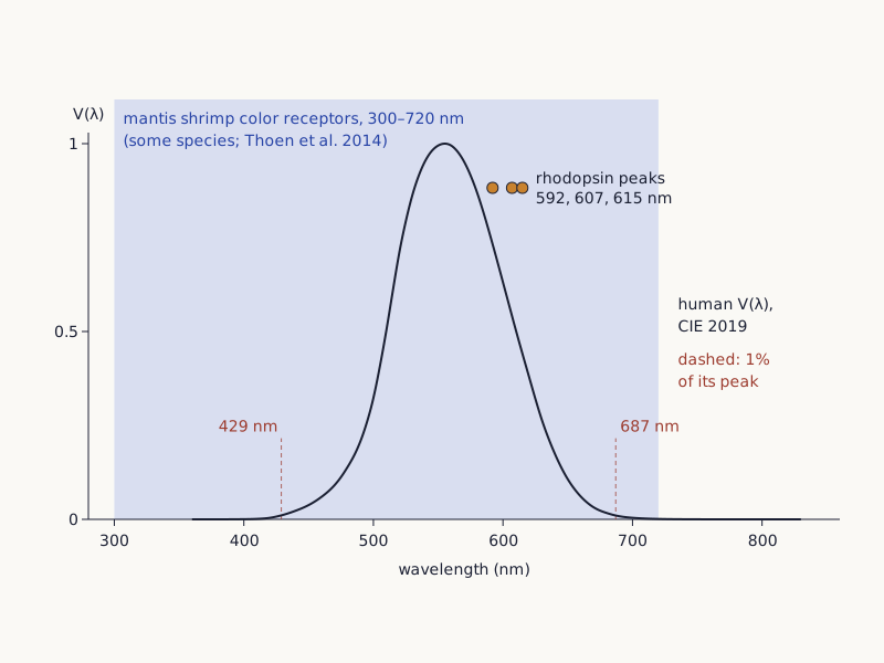
The shaded band in Figure 1 covers 300 to 720 nanometers, the span sampled by some mantis shrimp's color receptors. Some species have 12 photoreceptor types for color, yet behavioral tests found poor color discrimination (Thoen et al., 2014, abstract) [1]. Summarizing those tests, Wang and Marshall (2025) put the steps the animals needed at 15–25 nanometers, against 1–8 for humans [2]. Their 2025 gray-card tests found signs of opponent processing, so how the animals use 12 channels remains open [2].
Wide drawing on a small screen: swipe sideways, or open it full size.
Figure 1. The span sampled by some mantis shrimp's color receptors (300–720 nm, Thoen et al. 2014) over the CIE 2019 human photopic V(λ) curve; dots mark three Neogonodactylus oerstedii rhodopsin absorption peaks, not receptor peaks.
Chart: Tricks of Ear and Eye, CC BY-SA 4.0
Full credit line to copy
"Mantis shrimp color-receptor span and the human photopic V(λ) curve" by Tricks of Ear and Eye, https://tricksofearandeye.com/animal-senses/mantis-shrimp-eyes/, licensed under CC BY-SA 4.0, https://creativecommons.org/licenses/by-sa/4.0/
Data: CIE 2019, CIE spectral luminous efficiency for photopic vision, International Commission on Illumination (CIE), Vienna, AT, DOI: 10.25039/CIE.DS.dktna2s3, licensed under CC BY-SA 4.0, https://creativecommons.org/licenses/by-sa/4.0/; Thoen, How, Chiou & Marshall 2014, Science 343: 411–413 (span, from the abstract); Brouillon et al. 2026, Science Advances 12(42) (rhodopsin peaks)
How many receptors does a mantis shrimp eye have?
Three published counts describe mantis shrimp eyes, and each counts something different. Some species have 12 photoreceptor types for color, each tuned to a narrow band from deep ultraviolet to far red (Thoen et al., 2014, abstract) [1]. Cronin and colleagues (2014) count 16 receptor classes in all, in sets for ultraviolet, spatial, color and polarization vision, according to their abstract [3]. Streets, England and Marshall (2022) add the receptors for polarization and brightness to the 12 and reach 20 input channels [4].
| Count | What is counted | Source and how much was read |
|---|---|---|
| 12 | photoreceptor types for color, in some species | Thoen et al. 2014, abstract |
| 16 | functional receptor classes for ultraviolet, spatial, color and polarization vision | Cronin et al. 2014, abstract |
| 20 | input channels: the 12 color receptors plus receptors for polarization and intensity | Streets et al. 2022, full text |
In most shallow-water species, the midband, a strip of six special rows, runs across the middle of each eye. Rows 1 to 4 hold the color receptors. Each of their ommatidia (the repeating optical units of a compound eye) stacks three tiers of receptors, each with its own sensitivity, which gives the 12 classes. Rows 5 and 6 serve polarization vision, while the two halves of the eye above and below the midband pick up linear polarization and brightness (Wang & Marshall, 2025) [2].
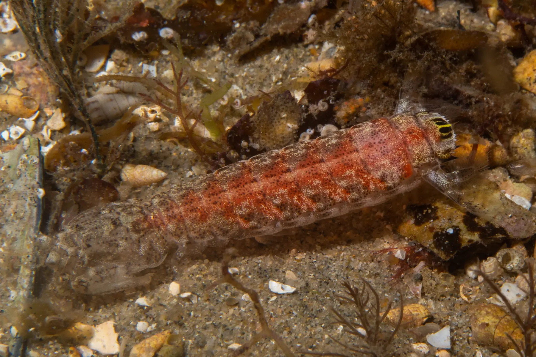
Figure 2. A slender mantis shrimp, Austrosquilla osculans, photographed in Australia in 2020; it is not the species used in the color tests on this page.
"Slender Mantis Shrimp (Austrosquilla osculans)" by Christopher Mark — https://commons.wikimedia.org/wiki/File:Austrosquilla_osculans_61399682.jpg — CC BY 4.0 (https://creativecommons.org/licenses/by/4.0/)
Most animals' color vision runs on two to four spectral sensitivities [4]. Wang and Marshall (2025) note that more than four add little to discrimination [2]. Pigeons have four classes of single cone, one with its pigment peak near 409 nm (Bowmaker et al., 1997, abstract) [5]. The pigeon vision entry draws the same line between counting cone types and measuring color vision.
How far past the human curve does the band reach?
Read Figure 1 from the left. The dark curve is V(λ), the CIE's standard luminous efficiency curve for human daylight (photopic) vision. Its table runs from 360 to 830 nm and peaks at 555 nm [6].
Computed for this page, the curve stays at or above 1% of its peak from 429 to 687 nm. The 1% cut is this page's choice. The mantis shrimp band starts 129 nm further toward the ultraviolet and ends 33 nm further toward the red.
This site calls a conversion like this a sensory exchange rate, which is this site's label, not a scientific term. Stated plainly, it compares the ends of two spans. Both spans are in nanometers, each has its own source, and the subtraction is in the range check file (CSV) (where the data come from and its license, JSON) Data: CIE 2019, CIE spectral luminous efficiency for photopic vision, International Commission on Illumination (CIE), Vienna, AT, DOI: 10.25039/CIE.DS.dktna2s3, licensed under CC BY-SA 4.0, https://creativecommons.org/licenses/by-sa/4.0/. V(λ) is a standard for measuring light, not a list of human receptors, so it is only a reference span here (V(λ) file (CSV) (where the data come from and its license, JSON) Data: CIE 2019, CIE spectral luminous efficiency for photopic vision, International Commission on Illumination (CIE), Vienna, AT, DOI: 10.25039/CIE.DS.dktna2s3, licensed under CC BY-SA 4.0, https://creativecommons.org/licenses/by-sa/4.0/).
The ends of the band vary by source. Thoen's abstract gives 300–720 nm [1], and Streets and colleagues write "from just below 300 nm to above 700 nm" [4]. Wang and Marshall give 300–750 nm [2], and Brouillon and colleagues give 300–700 nm in Neogonodactylus oerstedii [7]. The receptor facts file (CSV) (where the data come from and its license, JSON) Data: Thoen, How, Chiou & Marshall 2014, A different form of color vision in mantis shrimp (abstract); Cronin, Bok, Marshall & Caldwell 2014, Phil. Trans. R. Soc. B 369: 20130032 (abstract); Streets, England & Marshall 2022, Journal of Experimental Biology 225: jeb243699, CC BY 4.0; Wang & Marshall 2025, Journal of Experimental Biology 228: jeb247952, CC BY 4.0; Brouillon et al. 2026, Spectral tuning and signaling of diverse and far red–sensitive rhodopsins in mantis shrimp eyes, CC BY 4.0 lists all four.
Rhodopsin peaks are not receptor peaks
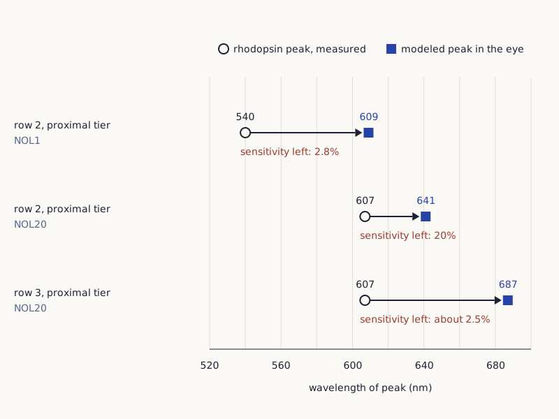
The three dots in Figure 1 mark a different kind of peak. Brouillon and colleagues (2026) made N. oerstedii rhodopsins, the light-absorbing pigment proteins of the receptors, in cultured cells. Three of them peak near or above 600 nm, at 615, 592 and 607 nm [7]. A peak measured this way belongs to a pigment, not a receptor: in rows 2 and 3, colored filters sit in front of the receptors, and the tiers above take part of the light first [7].
Wide drawing on a small screen: swipe sideways, or open it full size.
Figure 3. Two Neogonodactylus oerstedii rhodopsin peaks measured outside the eye, and the peaks Brouillon et al. (2026) model for them behind the filters of rows 2 and 3; the model leaves out signal processing and behavior.
Chart: Tricks of Ear and Eye, CC BY 4.0
Full credit line to copy
"Two rhodopsin peaks and their modeled peaks behind the filters, Neogonodactylus oerstedii" by Tricks of Ear and Eye, https://tricksofearandeye.com/animal-senses/mantis-shrimp-eyes/, licensed under CC BY 4.0, https://creativecommons.org/licenses/by/4.0/
Data: Brouillon et al. 2026, Spectral tuning and signaling of diverse and far red–sensitive rhodopsins in mantis shrimp eyes, CC BY 4.0
In their model (Figure 3), the rhodopsin that peaks at 607 nm on its own peaks at 687 nm behind the row 3 filter. Only about 2.5% of its unfiltered sensitivity is left [7]. The pigment Egyptian blue gives off light even further out, in a narrow infrared band near 910 nm (Seymour et al., 2020) [8], as the Egyptian blue entry explains.
Does 12 receptors mean 12 colors?
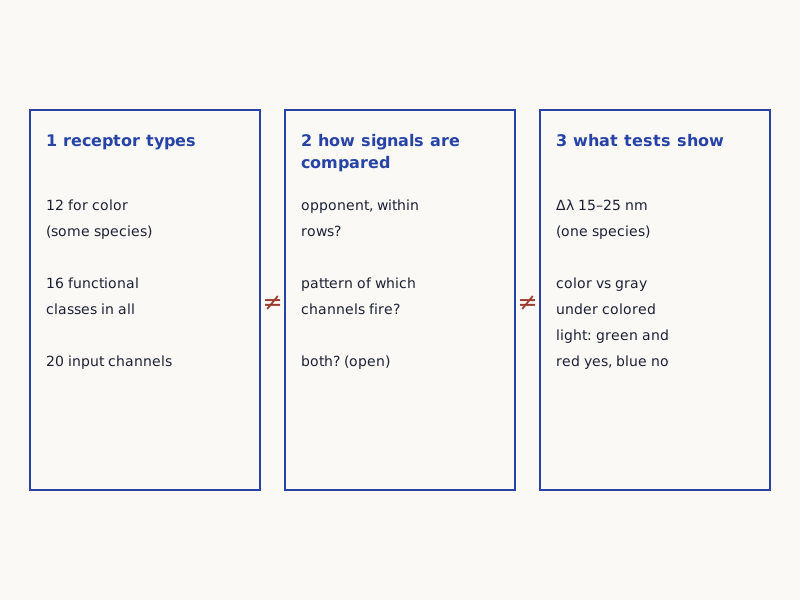
Figure 4 splits the question into three. How many receptor types an eye has is one question. How their signals are compared is another, and which colors an animal tells apart in a test is a third. Thoen and colleagues set out the puzzle in their abstract: three or four channels are enough for fine color discrimination, which leaves open why an eye would use 12 [1].
Wide drawing on a small screen: swipe sideways, or open it full size.
Figure 4. Three separate questions about mantis shrimp color vision: how many receptor types, how their signals are compared, and what behavior tests show.
Diagram: Tricks of Ear and Eye, CC BY 4.0
Full credit line to copy
"Receptor types, signal comparison and behavior: three questions" by Tricks of Ear and Eye, https://tricksofearandeye.com/animal-senses/mantis-shrimp-eyes/, licensed under CC BY 4.0, https://creativecommons.org/licenses/by/4.0/
Wang and Marshall (2025) describe two proposed schemes for the comparison step [2]. In the opponent scheme, each of rows 1 to 4 compares the signals of its own tiers, like four small two-receptor systems side by side. In the binning scheme, the color is read from which of the 12 channels responds most strongly, a pattern that Thoen and colleagues likened to the way the cochlea sorts sound [2]. Streets and colleagues suggested in 2022 that both could run at once [4].
The tests below measure which object a trained animal picks. They do not show what a color looks like to that animal. The bat echolocation entry meets the same gap between a measured signal and an animal's experience.
What did the color tests find?
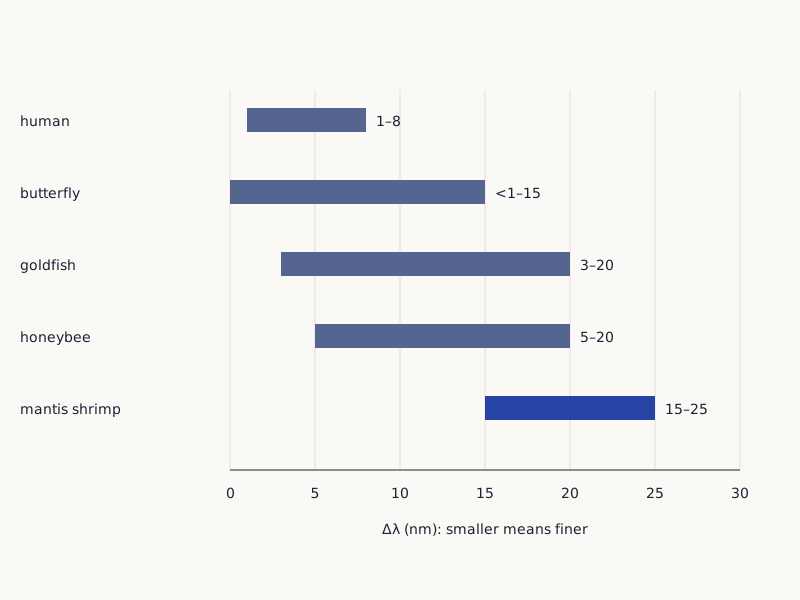
Compare the bottom bar of Figure 5 with the top one. Δλ is the gap in wavelength two lights need before an animal reliably tells them apart, so a shorter bar means finer discrimination.
In Thoen's tests, mantis shrimp needed 15–25 nm, as Wang and Marshall (2025) summarize them. The human figure they cite is 1–8 nm (De Valois and Jacobs, 1968) [2]. Honeybees, goldfish and butterflies have fewer receptor types, and all three reach finer steps than 15 nm at their best [2].
Wide drawing on a small screen: swipe sideways, or open it full size.
Figure 5. Smallest wavelength difference told apart in tests, as a range across the spectrum, for five animals as compiled by Wang and Marshall (2025).
Chart: Tricks of Ear and Eye, CC BY 4.0
Full credit line to copy
"Wavelength discrimination (Δλ) ranges for five animals" by Tricks of Ear and Eye, https://tricksofearandeye.com/animal-senses/mantis-shrimp-eyes/, licensed under CC BY 4.0, https://creativecommons.org/licenses/by/4.0/
Data: Wang & Marshall 2025, Journal of Experimental Biology 228: jeb247952, CC BY 4.0, citing De Valois & Jacobs 1968, Koshitaka et al. 2008, Neumeyer 1986, von Helversen 1972 and Thoen et al. 2014
| Animal | Δλ range (nm) | Original study, as cited by Wang & Marshall 2025 |
|---|---|---|
| human | 1–8 | De Valois & Jacobs 1968 |
| butterfly | below 1 to 15 | Koshitaka et al. 2008 |
| goldfish | 3–20 | Neumeyer 1986 |
| honeybee | 5–20 | von Helversen 1972 |
| mantis shrimp (Haptosquilla trispinosa) | 15–25 | Thoen et al. 2014 |
Thoen's abstract does not name the species tested. Wang and Marshall give the curve as that of Haptosquilla trispinosa, a reef mantis shrimp 2–3.5 cm long as an adult [2]. Streets and colleagues label the receptor curves from Thoen's study with the same species [4]. This page does not extend the result to every mantis shrimp.
According to their abstract, the poor scores ruled out color vision built on the usual opponent code. Thoen and colleagues proposed a different system: signals compared over time as the eyes scan, suited to recognizing a color rather than telling close ones apart [1]. Wang and Marshall read the same curve another way. It has four dips within 400–750 nm, and they suggest these line up with the spectral windows of rows 1 to 4, as an opponent scheme within each row would predict [2].
Whether mantis shrimp see more colors than people is not something these tests can show, because they do not record what any animal sees. On the narrower question they do answer, telling close wavelengths apart, the human figure is finer.
Can mantis shrimp tell a color from gray?
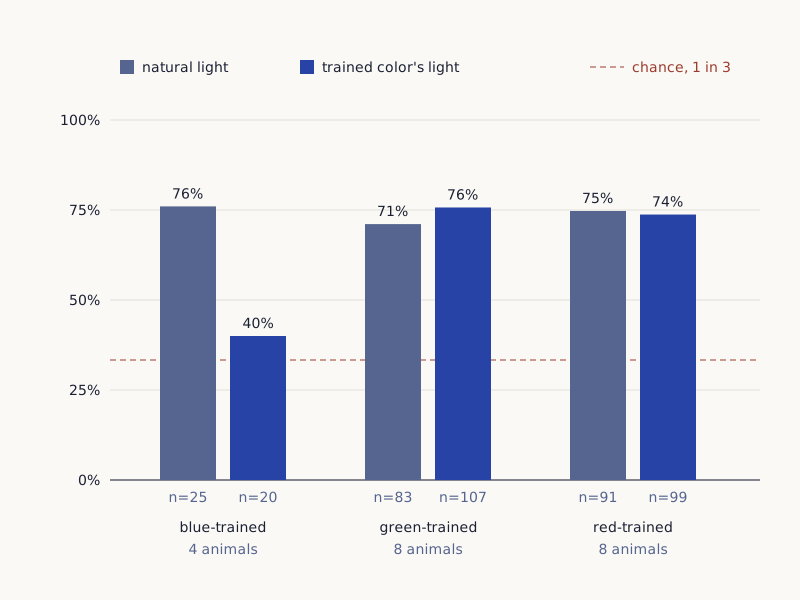
In Figure 6, find the one bar that falls close to the dashed chance line. In this gray-card test, each animal learned to grab one cable tie with a colored filter and to ignore two ties with gray filters. A random pick is right one time in three [2]. The animals, H. trispinosa from reefs at Lizard Island, were then tested in natural light and in tents lit with light of their trained color [2].
The colored light was the point. It cuts the light down to one band, so few color rows get much signal [2]. Only a comparison between receptor signals (opponent processing) should then still pick out the target, the authors reasoned, while binning alone should fail [2].
Wang and Marshall ran two experiments. In the first, green-trained animals scored 75.7% under green light, and red-trained animals 73.74% under red light. Blue-trained animals fell to 40%, not significantly different from chance [2]. The authors call this the first direct behavioral evidence of spectral opponency in mantis shrimp, and propose a hybrid of opponent and binning processing [2].
Wide drawing on a small screen: swipe sideways, or open it full size.
Figure 6. Gray-card success rates of Haptosquilla trispinosa under natural light and under light of the trained color, in the first of two experiments by Wang and Marshall (2025); chance is 1 in 3.
Chart: Tricks of Ear and Eye, CC BY 4.0
Full credit line to copy
"Gray-card test results, Haptosquilla trispinosa" by Tricks of Ear and Eye, https://tricksofearandeye.com/animal-senses/mantis-shrimp-eyes/, licensed under CC BY 4.0, https://creativecommons.org/licenses/by/4.0/
Data: Wang & Marshall 2025, Journal of Experimental Biology 228: jeb247952, CC BY 4.0
| Group | Natural light | Light of the trained color |
|---|---|---|
| blue-trained, 4 animals | 76% (25 trials) | 40% (20 trials) |
| green-trained, 8 animals | 71.08% (83 trials) | 75.7% (107 trials) |
| red-trained, 8 animals | 74.73% (91 trials) | 73.74% (99 trials) |
The groups are small. Of 69 animals trained, 20 performed reliably under natural light and were analyzed [2]. For the blue failure the authors offer two possible reasons and do not settle it: low contrast between their blue and gray cards, or a special role of blue signals in courtship [2]. All the numbers are in the color tests file (CSV) (where the data come from and its license, JSON) Data: Wang & Marshall 2025, Journal of Experimental Biology 228: jeb247952, CC BY 4.0; Streets, England & Marshall 2022, Journal of Experimental Biology 225: jeb243699, CC BY 4.0.
Streets and colleagues (2022) had already found that H. trispinosa could learn to tell several colors from grays, including a pale blue that the peacock mantis shrimp had failed with in an earlier test they describe [4]. Their own animals did worse the longer they were kept under artificial light. All learned the task in their first 10 weeks in captivity, but from weeks 11 to 20 their choices were no better than chance for any color [4]. The authors suggest this kind of change needs attention, and possibly a second look, in any color test on animals [4].
Why twelve, and what else do the eyes pick up?
The sources read for this page offer proposals, not an answer. Thoen and colleagues suggest recognition from a scan [1], which may allow faster processing at the cost of finer discrimination, Streets and colleagues note [4]. They cite an earlier idea that the narrow receptors help keep colors constant under water, an idea they call hypothetical. No known task in nature needs the full 12-way comparison, they write [4].
Wang and Marshall argue for the hybrid system instead [2]. It remains unclear, Streets and colleagues write, why the most complete set of spectral sensitivities known in any animal gives such poor color discrimination [4].
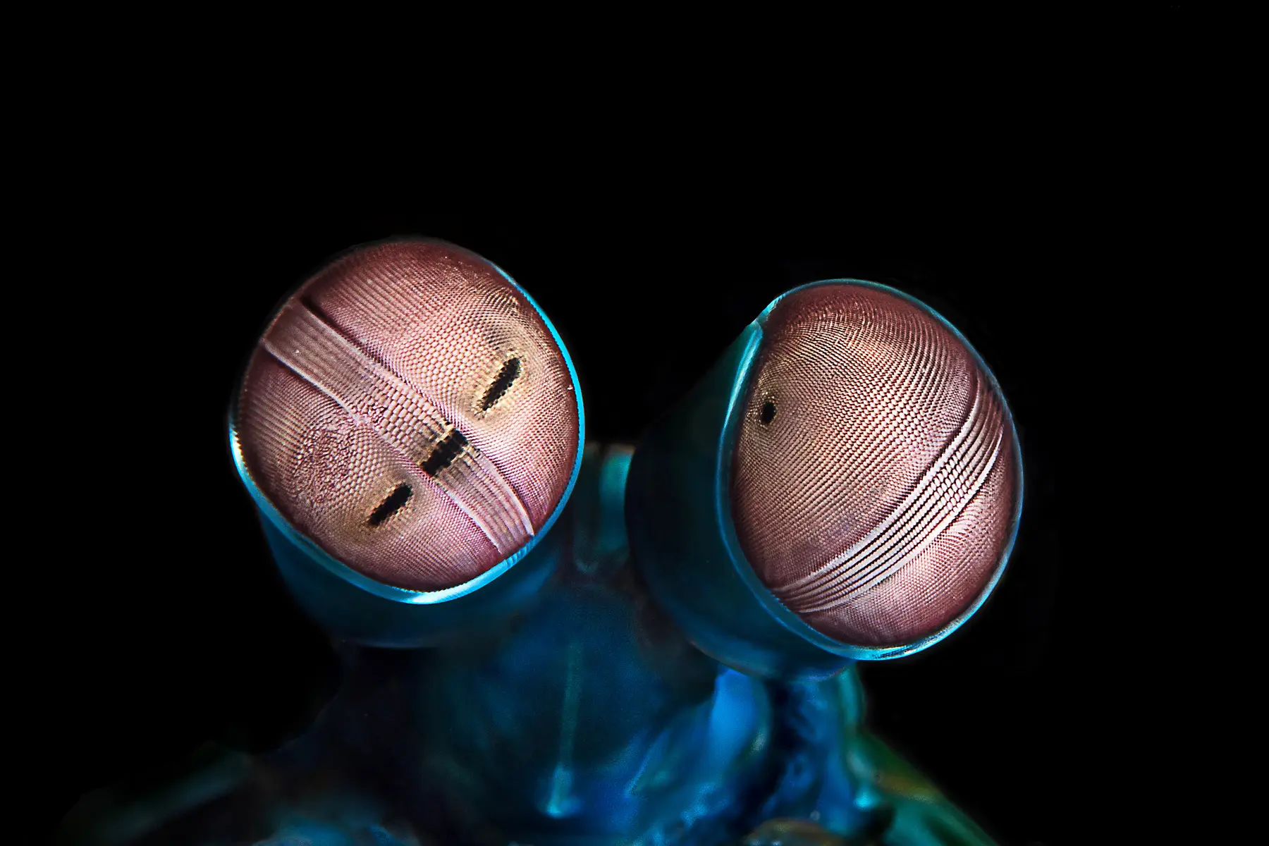
Figure 7. The eyes of a peacock mantis shrimp, Odontodactylus scyllarus, photographed at Réunion in 2016; it was not tested in the 2014, 2022 or 2025 studies.
"Odontodactylus scyllarus eyes" by Cédric Peneau — https://commons.wikimedia.org/wiki/File:Odontodactylus_scyllarus_eyes.jpg — CC BY-SA 4.0 (https://creativecommons.org/licenses/by-sa/4.0/)
Color is only part of the eye's work. Rows 5 and 6 of the midband detect circular (or elliptical) polarization, a property of light separate from its wavelength, at around 500 nm (Wang & Marshall, 2025) [2]. Cronin and colleagues write in their abstract that some ultraviolet receptors also act as filters that make circular polarization vision possible. Other filters, they add, extend these animals' vision deeper into the ultraviolet and further into the red [3].
Sources
- Thoen, H. H., How, M. J., Chiou, T.-H., & Marshall, J. (2014). A different form of color vision in mantis shrimp. Science 343(6169): 411–413. Open the sourceRead: abstract only
- Wang, C. J., & Marshall, N. J. (2025). Behavioural evidence of spectral opponent processing in the visual system of stomatopod crustaceans. Journal of Experimental Biology 228(1): jeb247952. Open the sourceRead: full text
- Cronin, T. W., Bok, M. J., Marshall, N. J., & Caldwell, R. L. (2014). Filtering and polychromatic vision in mantis shrimps: themes in visible and ultraviolet vision. Philosophical Transactions of the Royal Society B 369(1636): 20130032. Open the sourceRead: abstract only
- Streets, A., England, H., & Marshall, J. (2022). Colour vision in stomatopod crustaceans: more questions than answers. Journal of Experimental Biology 225(6): jeb243699. Open the sourceRead: full textText license: CC BY 4.0
- Bowmaker, J. K., Heath, L. A., Wilkie, S. E., & Hunt, D. M. (1997). Visual pigments and oil droplets from six classes of photoreceptor in the retinas of birds. Vision Research 37(16): 2183–2194. Open the sourceRead: abstract only
- CIE (2019). CIE spectral luminous efficiency for photopic vision [data set]. International Commission on Illumination (CIE), Vienna, AT. Open the sourceRead: full textText license: CC BY-SA 4.0
- Brouillon, C., Pushkarev, A., Liénard, M., Hwang, S., Munhoven, A., McDowell, R., Lucas, R., Porter, M., Sun, H., & Hegemann, P. (2026). Spectral tuning and signaling of diverse and far red–sensitive rhodopsins in mantis shrimp eyes. Science Advances 12(42). Open the sourceRead: full text
- Seymour, L. M., Nicola, M., Kessler, M. I., Yost, C. L., Bazzacco, A., Marello, A., Ferraris, E., Gobetto, R., & Masic, A. (2020). On the production of ancient Egyptian blue: Multi-modal characterization and micron-scale luminescence mapping. PLoS ONE 15(11): e0242549. Open the sourceRead: full textText license: CC BY 4.0
Sense check:
One step, under a minute
Find where the human curve in Figure 1 fades out at the short-wavelength end, then read how far below it the mantis shrimp band starts. Next: Pigeon vision · Egyptian blue
Related entries
Bat Echolocation: In a Study, Search Calls Peaked at 25+ kHz
Animal Senses
In field recordings of 11 bat species in Panama, the strongest call frequency was above 20 kHz in every one. How loud the calls were, and how echoes give range.
Image credit
"Greater Bulldog Bat, Noctilio leporinus" by Susan Ellis, Bugwood.org — https://commons.wikimedia.org/wiki/File:Captive_Noctilio_leporinus.jpg — CC BY 3.0 (https://creativecommons.org/licenses/by/3.0/us/)
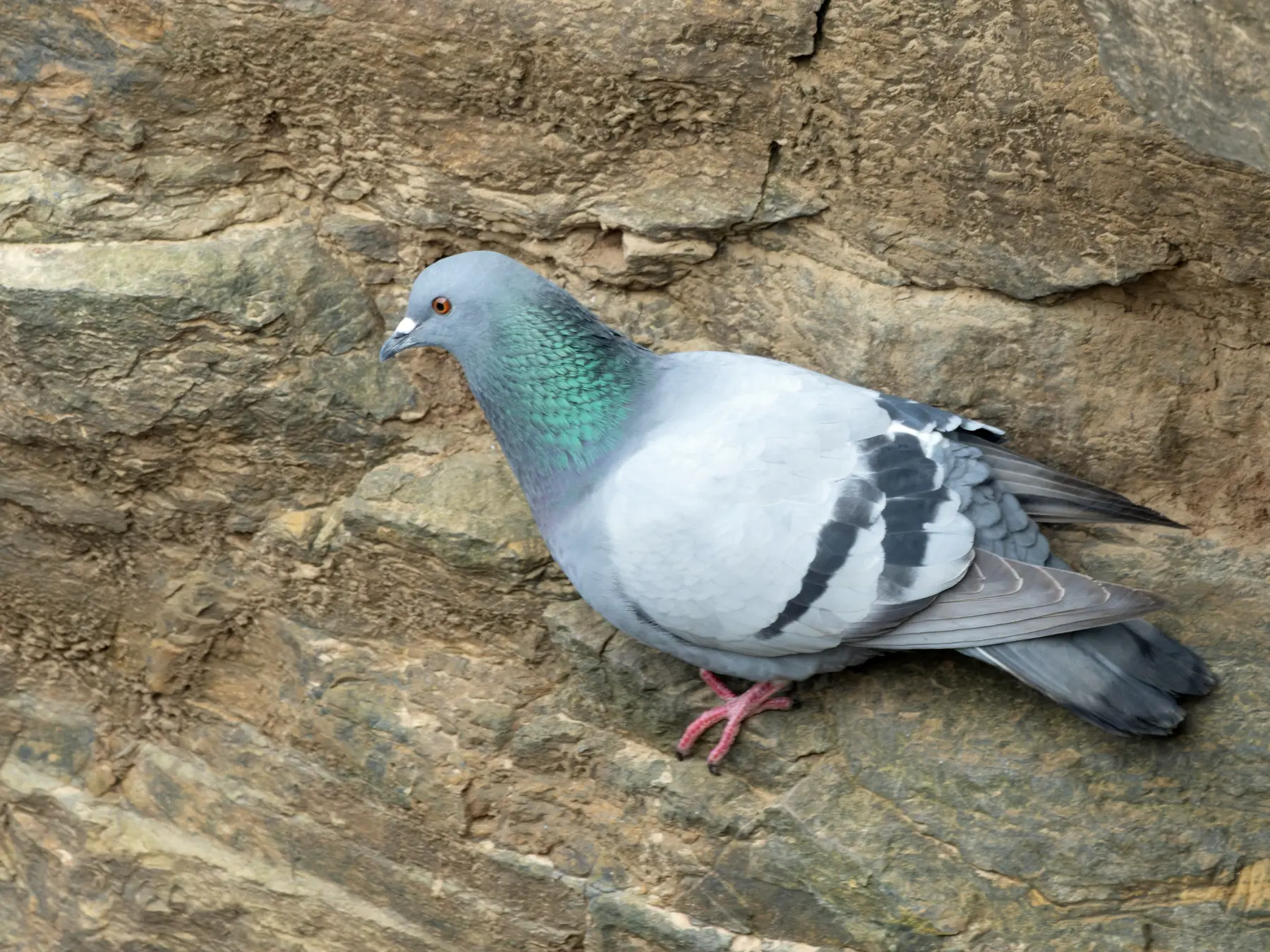
Pigeon Vision: Most of What It Sees, It Sees With One Eye
Animal Senses
Pigeon vision: both eyes share about 20° in front; about 40° at the back is blind (2024 summary; values differ). Shortest cone: about 409 nm (1997 abstract).
Image credit
"Columba livia (Rock Dove, wild), Duncansby Head, Caithness, Scotland" by william_stephens — https://commons.wikimedia.org/wiki/File:Columba_livia_(Rock_Dove,_wild),_Duncansby_Head,_Caithness,_Scotland_1.jpg — CC BY 4.0 (https://creativecommons.org/licenses/by/4.0/)
Cite this page
Tricks of Ear and Eye. "Mantis Shrimp Eyes: A Tested Species Told Hues Apart Poorly." Tricks of Ear and Eye. Published 2026-10-30. https://tricksofearandeye.com/animal-senses/mantis-shrimp-eyes/. Accessed on the date you read it.